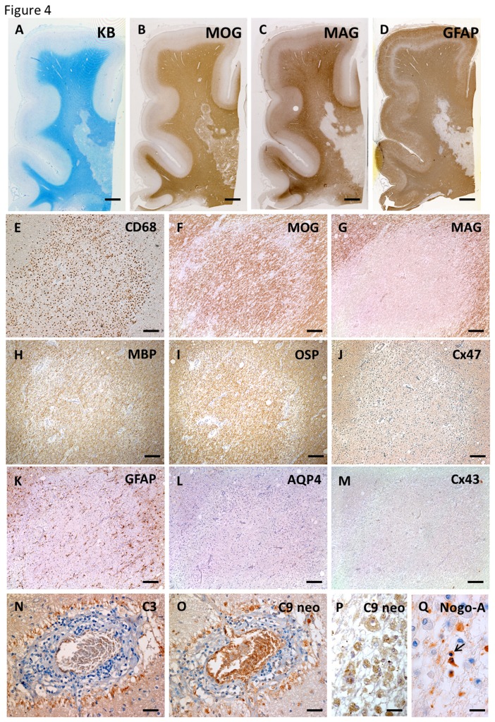
Figure 4. Distal oligodendrogliopathy and astrocytopathy in anti-AQP4 antibody-seropositive NMO (case NMO-10).
In active lesions of the cerebral white matter, KB staining and MOG immunostaining show remaining myelin (A, B). Patterns of preferential MAG loss and marked loss of GFAP immunoreactivity are seen (C, D). Higher magnification reveals sharply demarcated, prominent MAG loss in this lesion with infiltration of numerous CD68-positive macrophages, whereas immunoreactivity for MOG, MBP and OSP is preserved in the lesion (E–I). Immunoreactivity for Cx47 is diminished compared with non-affected white matter (J). Complete loss of AQP4 and Cx43 in degenerative, GFAP-positive astrocytes (K–M) and complement deposition are observed around blood vessels with perivascular cell cuffing (N, O). Complement components are present within foamy macrophages in this lesion (P). Nogo-A-positive oligodendrocytes are markedly decreased in this lesion and some remaining oligodendrocytes show nuclear condensation, suggesting apoptotic changes (Q). This lesion is classified as pattern A for Cx43 and pattern B for Cx47/Cx32. Scale Bar = 4 mm (A–D); 200 µm (E–M); 50 µm (N, O); 20 µm (P, Q).

