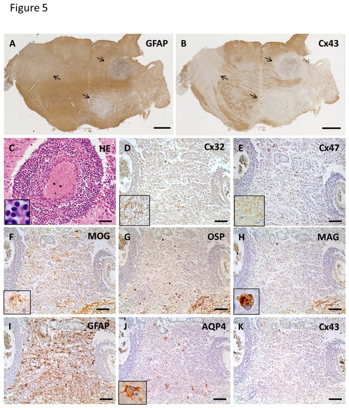
Figure 5. Cx43 and AQP4 astrocytopathy in active lesions of MS (case MS-3).
Low magnification view of GFAP (A) and Cx43 (B) immunostaining in the pons. Immunoreactivity for Cx43 is markedly diminished in multiple lesions covered by GFAP-positive astrocytes (A, B, arrows). Massive perivascular cuffing, mostly consisting of lymphocytes, is observed in these lesions (C). Immunoreactivities for Cx32 and Cx47 are decreased in these lesions (D, E) compared with non-affected white matter (D, E, insert). Loss of MAG compared with MOG and OSP is prominent (F–H). Infiltration of macrophages phagocytosing myelin debris, which are immunopositive for myelin proteins (F, H, insert). Numerous GFAP-positive hypertrophic astrocytes exist in perivascular areas and parenchyma of these lesions (I). Patchy loss of AQP4 and diffuse loss of Cx43 in the center and periphery of lesions (J, K). Some hypertrophic astrocytes demonstrate membranous staining for AQP4 (J, insert). This lesion is classified as pattern A for Cx43 and pattern B for Cx47/Cx32. Scale Bar = 4 mm (A, B); 50 µm (C); 100 µm (D–K).

