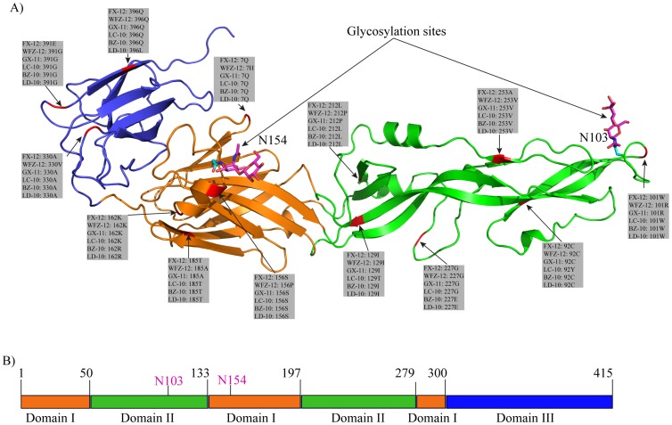
Figure 2. Modeled structure and domain architecture of envelope protein of FX-2012 strain.
A) Cartoon scheme view of envelope protein structure. Sites containing amino acid changes were colored red and all these changes were listed in gray boxes. The side-chains of glycosylated residues were colored cyan. For illustration purpose, a β-D-Manp-(1-4)-β-D-GlcpNAc (colored magenta) was added to the glycosylation sites. B). Domain diagram of envelope protein. Domain boundaries were labeled (black) on top of the diagram. The two glycosylation sites were labeled magenta. Domain boundaries were predicted based on domain architecture of Japanese encephalitis virus. Domain colors: orange, domain I; green, domain II; blue, domain III.

