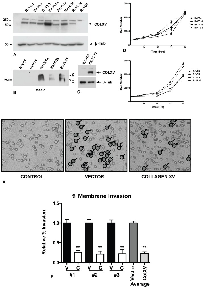
Figure 1. Collagen XV inhibits invasion of BxPC cells though a collagen I-coated membrane.
A) Stable expression of collagen XV in BxPC-3 and S2-013 pancreatic adenocarcinoma cells. Western blot probed with anti-COLXV antibody or anti-ß-tubulin loading control. Clones Bx15.1-15.40 express variable levels of COLXV. Also shown is one vector control clone (BxVC1). Note: the anti-COLXV antibody cross-reacts with an irrelevant higher MW protein that is seen in many cell types, irrespective of COLXV expression. B) The COLXV is secreted from BxPC-3 cells into the cell culture media. C) COLXV expression in a representative stable clone of S2-013 cells (S2.15.10), also shown is a vector control (S2.VC3). D) Growth rates of 4 BxPC-3 vector control clones and 4 COLXV-expressing clones are independent of COLXV expression. E, F) Invasion assay: E) vector (BxVC1, BxVC3) or COLXV (Bx15.14 and Bx15.23) cells plated on a membrane coated with COLI. After 72 hr cells invading through the membrane were stained, visualized and counted at 40X magnification. Random fields were selected from three independent experiments representing n = 16 vector plates (BxVC1, BxVC3) and n = 15 COLXV plates (Bx15.14 and Bx15.23). F) The average of each independent experiment is shown #1, #2, #3, (Left, V = vector control; C = COLXV) and the cumulative average of all triplicates (right).

