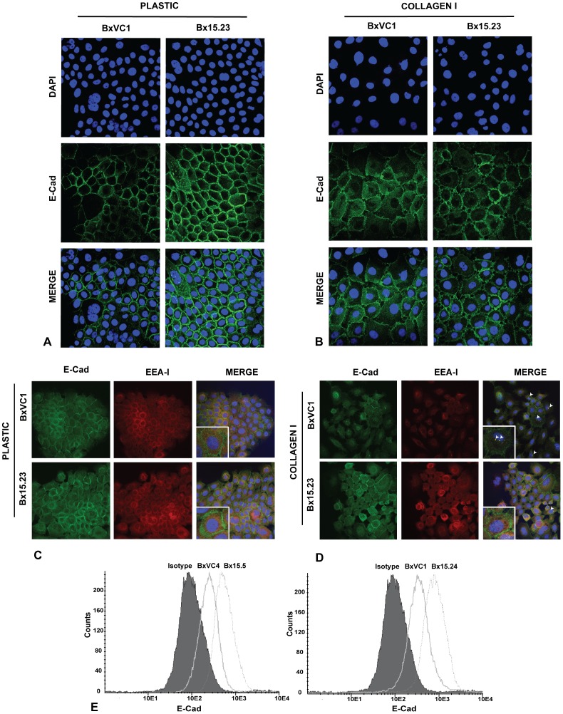
Figure 3. E-Cadherin is stabilized at the cell periphery in collagen XV expressing cells.
Confocal microscopy with an antibody specific for the extracellular domain of E-Cad (green) and nuclei stained with DAPI (blue). BxVC1 vector clone and Bx15.23 COLXV clone grown on plastic A) or COLI B). E-Cad is most abundant at the cell surface in both clones on plastic. On COLI, E-Cad moves from the cell periphery into the cytoplasm in BxVC1, but this redistribution is inhibited in the presence of COLXV (BX15.23). C) EEA1 (red) is found in the endoplasmic reticulum (ER)/Golgi zone of the cells grown on plastic, while E-Cad is at the cell periphery. D) After relocation of E-Cad on COLI, EEA1 colocalizes with E-Cad (white arrowheads) in BxVC1 cells but not Bx15.23 cells. Images are representative of several clones. E) Flow cytometry after staining cells with an E-Cad antibody shows increased cell-surface expression of E-Cad in cells with COLXV (Bx15.5 and 15.24) in comparison to vector controls (BxVC4 and BxVC1). All experiments performed a minimum of 3 times with consistent results.

