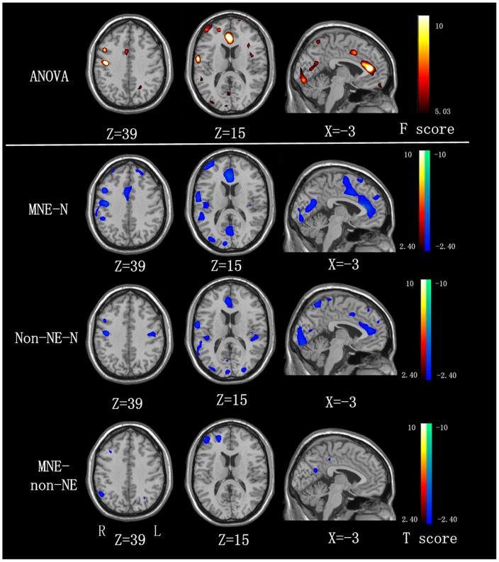
Figure 2. ReHo differences among MNE, non-NE, and healthy controls (P<0.05, AlphaSim corrected).
Compared with the healthy controls, MNE patients show decreased ReHo in the bilateral SMA, ACC, PCC, MOG, right insula, cuneus, MFG, IPL, STG, PCu, and left PreCG, PoCG, and non-NE patients show decreased ReHo in the bilateral ACC, cuneus, precuneus, STG, PreCG, left PoCG, MOG and right MFG, and Compared with the non-NE patients, the MNE patients show decreased ReHo in the right IPL, MFG and left precuneus. MNE = minimal nephro-encephalopathy; non-NE = non-nephro-encephalopathy; ReHo = regional homogeneity; IPL = inferior parietal lobe; PCu = precuneus; ACC = anterior cingulate cortex; PCC = posterior cingulate cortex; SMA = supplementary motor area; PreCG = precentral gyrus; PoCG = postcentral gyrus; SFG = superior frontal gyrus; MFG = medial frontal gyrus; STG = superior temporal gyrus; MOG = medial occipital gyrus.

