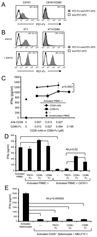
Figure 5. CD80-Fc restores PBMC activation by neutralizing PDL1-PD1 suppression and by costimulating through CD28.
(A) Expression of CD80 prevents binding of PD1 to PDL1+ tumor cells. C8161 and C8161/CD80 human tumor cells were incubated with or without recombinant human PD1-Fc fusion protein followed by staining with mAb to PD1 and analyzed by flow cytometry (top panel). (B) 4T1 and 4T1/CD80 mouse tumor cells were cultured with IFNγ for 48hrs, followed by incubation ± unlabeled PDL1 mAb 43H12, which disrupts PDL1-CD80 interactions. Tumor cells were subsequently incubated with recombinant mouse PD1-Fc followed by staining with mAb to PD1, and analyzed by flow cytometry (bottom panels). (C) CD80-Fc has less costimulatory activity than antibodies to CD28. Healthy donor PBMC were activated with PHA and cultured with CD80-Fc or antibodies to CD28. IFNγ production was measured by ELISA. Molar concentration for antibody was ½ the molar concentration of CD80-Fc to compensate for each antibody molecule binding two molecules of CD28 vs. each CD80-Fc molecule binding only one molecule of CD28. (D) CD80-Fc restores PBMC activation more effectively than CD86-Fc. PHA-activated PBMC from healthy donors were co-cultured with or without C8161 human tumor cells in the presence or absence of CD80-Fc, CD86-Fc, or TROY-Fc. IFNγ production was measured by ELISA. (E) Splenocytes from CD28−/− mice were activated with PMA and ionomycin and co-cultured with MELF10 mouse tumor cells ± CD80-Fc, CD86-Fc, or TROY-Fc. Data are representative of 3, 3, 3, and 3 independent experiments for (A), (B), (C), and (D) respectively.

