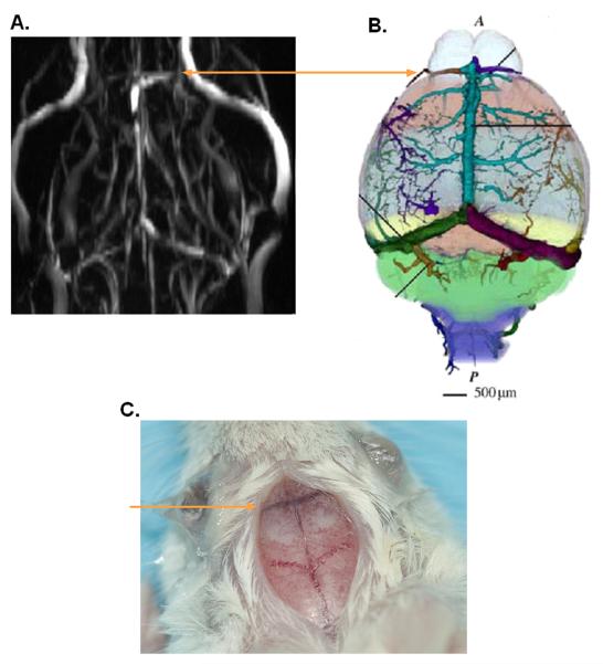
Fig. 1. Presence of the rostral rhinal vein on the surface of a mouse brain.
A, Angiogram of a non-tumor bearing GFAP-CreER™;Ptenflox/flox;Tp53flox/flox;Rb1flox/flox mouse showing a large vein across the olfactory bulb/frontal lobe border. B, Diagrammatic illustration of veins on mouse brain surface illustrating the rostral rhinal vein on the border between the olfactory bulb and the mouse brain frontal lobe. Reproduced with permission from16. C, Brain surface of a GFAPCreER™;Ptenflox/flox;Tp53flox/flox;Rb1flox/flox mouse. Lines indicate location of the rostral rhinal vein.

