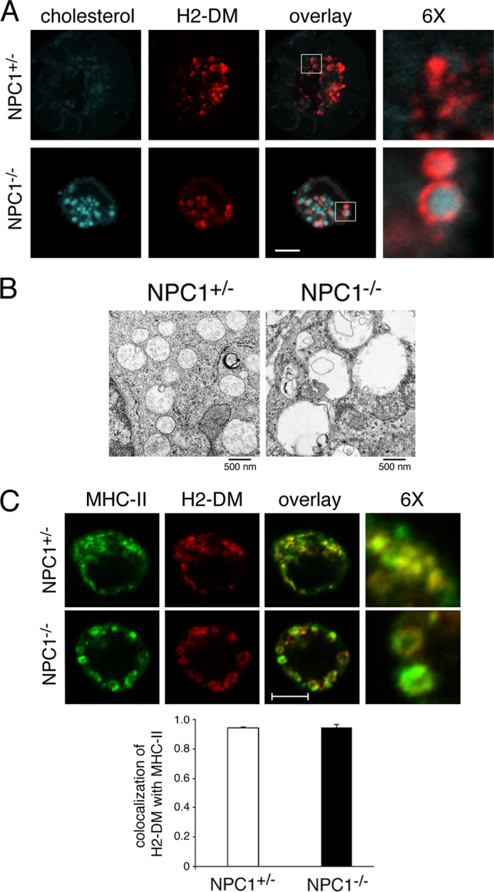
FIGURE 1.

MVB ultrastructure and MHC-II localization are disturbed in lipid-overloaded MVBs. A, control or NPC1−/− DCs were harvested after 7 days of growth in medium and attached to polylysine-coated coverslips prior to fixation, permeabilization, and staining with filipin to detect unesterified cholesterol (blue) and a mAb recognizing H2-DM (red). 6× magnified images of the indicated regions of the merged image are shown. Scale bar represents 5 μm. B, DCs were fixed and sectioned for electron microscopy. Scale bar represents 500 nm. C, immature DCs were fixed, permeabilized, and stained with mAbs recognizing MHC-II (M5/114, green) or H2-DM (red). 6× magnified images of the indicated regions of the merged image are shown. Scale bar represents 5 μm. The extent of colocalization of H2-DM+ red pixels with MHC-II+ green pixels was determined using software provided with the LSM510 confocal microscope.
