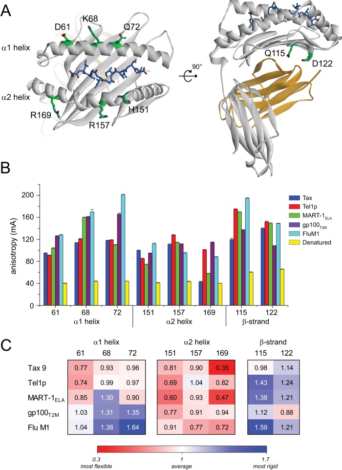
FIGURE 2.
Fluorescence anisotropy indicates a peptide dependence to HLA-A2 molecular flexibility. A, location of the fluorescently labeled sites in the HLA-A2 molecule. In the structures of the wild-type protein, the sites chosen for labeling were all solvent-exposed, polar residues whose side chains made no contacts to the peptide. B, anisotropy values (in mA, or anisotropy reading × 10−3) measured at each site for the various labeled peptide·HLA-A2 complexes. Values plotted are the means and standard deviations of 50 measurements on two independently prepared samples. C, anisotropy values in panel B recast as the percentage of deviation from the average. Higher numbers and blue shading reflect less than average flexibility. Lower numbers and red shading reflect greater than average flexibility.

