
FIGURE 3.
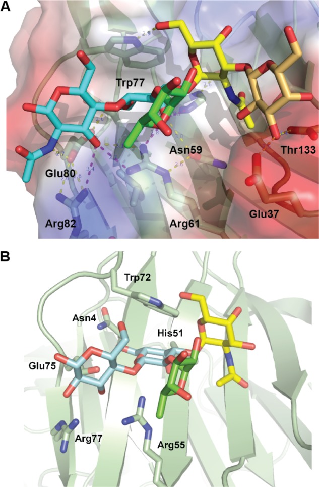
Recognition of blood group A type 2 oligosaccharides by CvGal1 and CGL2. A, A2 blood antigen docked at the binding pocket of the CvGal1 model of the first CRD, using the observed common N-acetyllactosamine disaccharide bound to the template. CvGal1-binding site is shown as semi-transparent solvent-accessible surface colored by its vacuum electrostatic potential (positive in blue to negative in red). The schematic model of the protein is visible across the surface showing the interacting residues in a stick representation. H-bonds recognizing hydroxyl groups of the A2 antigen are displayed as dashed lines with their distances (in Å) between heavy atoms indicated. B, A2 bound to CGL2; PDB code 1UFL. The carbon atoms of the N-acetyllactosamine moiety are colored in cyan, the α(1–3)GalNAc in yellow, and α(1,2)-Fuc in green. All nitrogen atoms are colored in blue. See also supplemental Fig. S2.