
FIGURE 4.
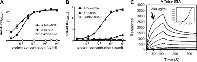
Binding of rCvGal1 and anti-A monoclonal antibodies to neoglycoproteins. A and B, ELISA: blood group A tetrasaccharide-BSA (tetra-BSA), blood group A trisaccharide-BSA (tri-BSA), or N-acetylgalactosamine-BSA (GalNAc-BSA) were delivered at the concentrations indicated (serial dilution starting from 10 μg/ml, 100 μl/well) into 96-well plates and incubated overnight. After blocking and washing the plates, the binding of mouse monoclonal anti-A antibodies (1:2000) (A) and CvGal1 (0.2 μg/ml) (B) was assessed. C, SPR measurements: blood group A tetrasaccharide-BSA, blood group A trisaccharide-BSA, or GalNAc-BSA were immobilized on CM5 chips, and binding of rCvGal1 was assessed by SPR flowing through rCvGal1 in 2-fold serial dilutions (starting from 200 μg/ml) as analyte. Sensorgrams and the 1:1 affinity curve (inset) for the binding of rCvGal1 onto blood group A tetrasaccharide-BSA are shown. Negligible responses were observed on sensorgrams for the other two neoglycoproteins, blood group A trisaccharide-BSA and GalNAc-BSA (data not shown).