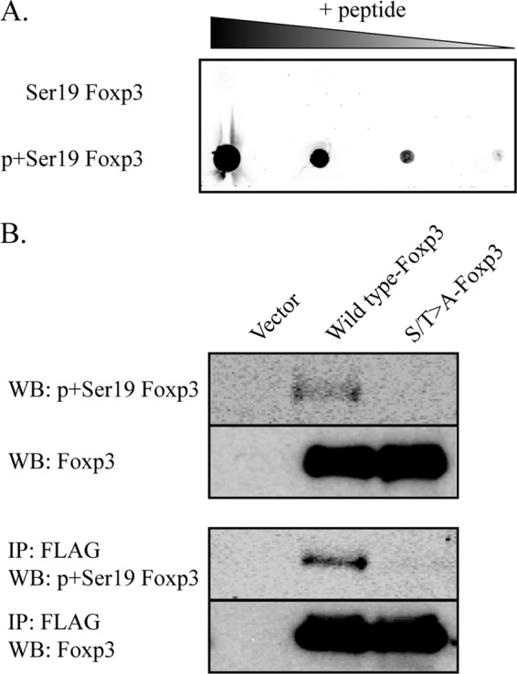
FIGURE 3.

Foxp3 is phosphorylated at CDK motifs in vivo. A, Foxp3 peptide (aa 12–24) containing either unmodified or phosphorylated serine 19 was serially diluted and used for a dot blot assay. Membranes containing diluted peptides were probed with p+Ser-19-Foxp3 antibody. B, HEK293 cells were transfected with vector control, wild type Foxp3, or S/T→A-Foxp3 mutant. Whole cell extracts were prepared at 48 h after transfection. FLAG immunoprecipitation (IP) and Western blotting (WB) were performed as described under “Experimental Procedures.” Phosphorylated and total Foxp3 is shown. Data are representative of three separate experiments.
