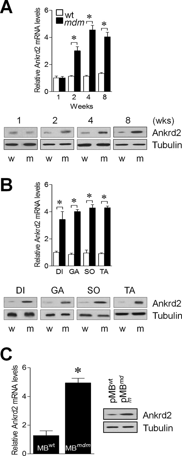
FIGURE 2.

Skeletal muscles of mdm mice over express ANKRD2. Hind limb skeletal muscles were excised from WT and mdm mice, and ANKRD2 expression was determined by real-time RT-PCR and Western blot methods (A). diaphragm (DI), gastrocnemius (GA), soleus (SO), tibialis anterior (TA), and primary myoblasts (pMB) were isolated from the hind limb skeletal muscles of WT and mdm mice, and ANKRD2 expression was determined by real-time RT-PCR and Western blot (B and C). w and m indicate wild-type and mdm, respectively. Gel images are representative of three separate experiments. Each error bar indicates mean ± S.E. (n = 3). *, p < 0.05. Wks., weeks.
