
FIGURE 1.
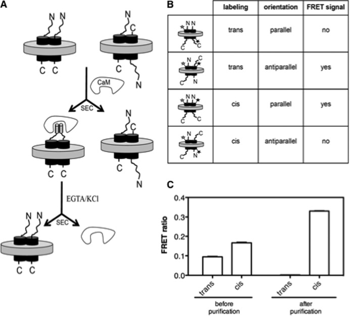
Parallel dimer purification. A, schematic representation of the PGD tag-based purification method. The tag is fused to the N terminus of the receptor and is represented schematically as a cylinder in the presence of CaM to account for its helical structure upon binding to CaM. Parallel dimers were separated from non-parallel ones using SEC. Another possibility would be to consider a CaM affinity-based method using calmodulin immobilized on a chromatographic matrix (CaM-Sepharose 4B). B, schematic representation and putative associated interprotomer FRET signal of the different species considered. The white star represents the fluorescence donor and the black one the acceptor. C, FRET ratio measured between Alexa Fluor 488 and Alexa Fluor 568-labeled BLT1 protomers reconstituted in lipid nanodiscs before and after purification using the PGD tag. Results are given as mean ± S.D. from three different experiments.