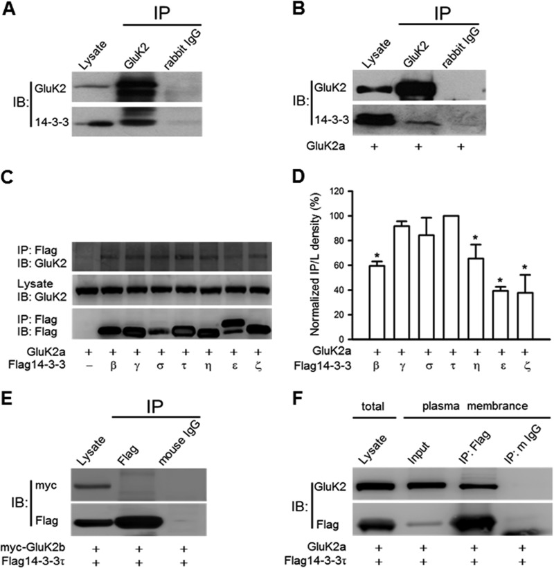
FIGURE 1.
14-3-3 proteins associate with GluK2a-containing KARs. A, GluK2 subunits coimmunoprecipitate with 14-3-3 proteins from rat hippocampal lysates. B, GluK2 coimmunoprecipitates with endogenous 14-3-3 proteins in HEK293 cells transfected with GluK2a cDNA. The anti-pan 14-3-3 antibody was used for immunoblotting (IB) in A and B; images shown are representative Western blots from three independent experiments. C, coimmunoprecipitation of GluK2a and different FLAG14-3-3 isoforms. The identities of the transfected cDNAs are indicated below each lane. The exogenously expressed 14-3-3 proteins were immunoprecipitated (IP) with the FLAG antibody. D, normalized levels of coimmunoprecipitation between GluK2a and various 14-3-3 isoforms. These values were determined by measuring the relative intensity of immunoprecipitated bands and their corresponding lysate (L) bands on Western blots and then normalized to and compared with that of 14-3-3τ (n = 3, *, p < 0.05). E, no interaction between GluK2b and 14-3-3τ is detected by coimmunoprecipitation in cotransfected HEK293 cells. F, GluK2a and FLAG14-3-3τ coimmunoprecipitate in the crude plasma membrane fraction prepared from cotransfected HEK293 cells. Image shown is representative Western blots from three independent experiments. Data represent mean ± S.E.; IB, antibody used for immunoblot analysis; IP, antibody used for immunoprecipitation.

