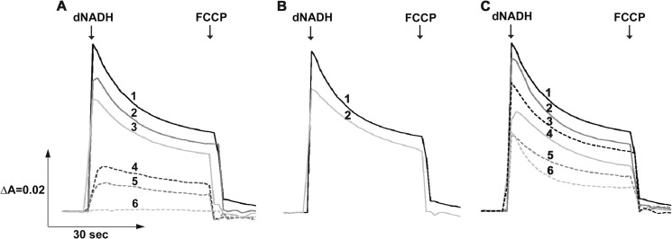
FIGURE 5.
Detection of the membrane potential generated by dNADH oxidation in NDH-1 mutants. The potential changes (ΔΨ) of E. coli membrane samples were monitored by the absorbance changes of oxonol VI at 630–603 nm at 30 °C. The first arrow indicates addition of dNADH, whereas the second arrow indicates addition of FCCP. Representative traces from different groups of mutants: A, conserved charged residue mutants: 1, WT (or NKO-rev, NE133A, NE133A/KKO-rev, NK217C, and NK247R); 2, NK217A (or NK217R); 3, NK247A (or NK395R); 4, LR175A (or LK342A and LKO); 5, NK395A (or ME407A and NE133A/KE72A); 6, NKO (or NE133A/KKO); B, conserved proline mutants: 1, WT (or NP387G); 2, NP222A (or NP387A, MP239A, MP399A, LP234A, and LP390A); and C, structural element residues: 1, WT; 2, NK158A (or NH224A and NV469A); 3, NAla481stop; 4, NK158R; 5, NIle475stop; 6, NVal469stop.

