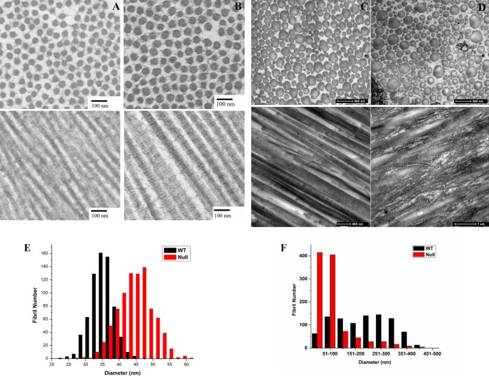
FIGURE 9.
Electron micrographs of tendon fibrils of wild type and P3H1 null mice. A and B, tendon fibrils of wild type (A) and P3H1 null (B) mice at P5 in cross sections and longitudinal sections and the distribution of measured diameters from the cross sections underneath. The length of the bars in the micrographs corresponds to 100 nm. C and D, tendon fibrils of adult wild type (C) and P3H1 null mice (D) in cross sections and longitudinal sections. The length of the bars corresponds to 500 nm. E and F, the diameter distribution from cross sections is given for P5 (E) and for adult mice (F). Panels C and D are similar to fields published previously, and panel F is taken from Fig. 2E in Ref. 13.

