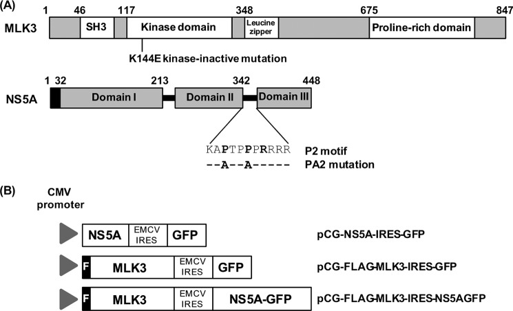
FIGURE 1.
A, molecular organization of MLK3 and HCV NS5A. Schematic illustration of the domain structure of the two proteins. Amino acid residue numbers indicated. The sequence of the P2 motif and the mutations introduced to generate the PA2 mutant are illustrated. The black box on NS5A is the N-terminal membrane-associating amphipathic helix. B, structure of bicistronic vectors used for transfection in this study. The construction of these vectors is detailed in the “Experimental Procedures.”

