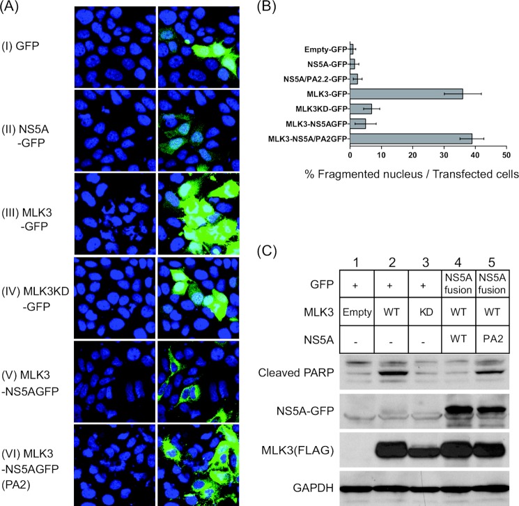
FIGURE 3.
Exogenous MLK3 expression induces apoptosis. A, Huh7 cells were seeded on coverslips and transfected with the indicated expression vectors. 24 h after transfection, cells were fixed and stained with DAPI. Fluorescence microscopy was performed to visualize transfected cells (GFP fluorescence) and the nucleus (DAPI). B, multiple fields were counted (n = 4). GFP-positive cells, more than 60 cells for each sample point, were counted as transfected cells and the percentage of GFP-positive cells showing fragmented nuclei are presented. C, Huh7 cells were transfected with the indicated plasmid vectors. Lysates were subjected to Western blotting to detect caspase-cleaved PARP (as a marker of apoptosis), as well as NS5A-GFP and FLAG-MLK3.

