FIGURE 6.
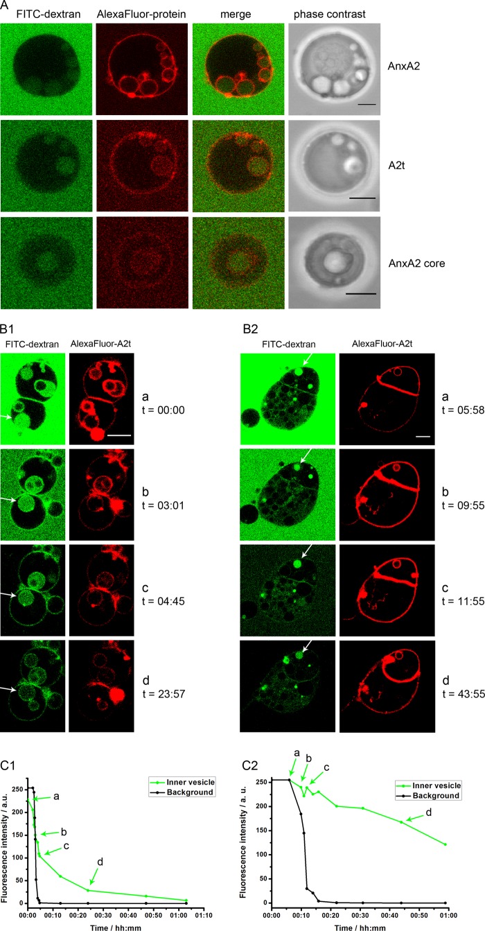
Characterization of ILVs induced by AnxA2 and derivatives. A, unlabeled GUVs were kept in 1 mg/ml FITC-dextran (Mr 150) and incubated in the presence of 250 μm Ca2+ together with AlexaFluor® 568-labeled AnxA2 (91.3 nm), A2t (102 nm), or the AnxA2 core domain Δ32 (1371 nm). Note that the formation of ILVs is accompanied by the uptake of FITC-dextran. B, unlabeled GUVs were placed into a flow chamber and incubated with AlexaFluor® 568-labeled A2t (100 nm) in PBS containing 1 mg/ml FITC-dextran (Mr 150) and 250 μm Ca2+ for 30 min at 25 °C. Subsequently, the mixture was washed with PBS containing 250 μm Ca2+ without FITC-dextran, and decline of FITC fluorescence was recorded by on-line microscopy. C, fluorescence intensity profiles of ILVs is depicted by a white arrow in B with C1 corresponding to B1 and C2 corresponding to B2. Time point 0 denotes the start of the washing. The ILV fluorescence was measured in the center of each vesicle, and the background intensity was measured outside of the vesicles. Bars, 5 μm. a.u., absorbance units.

