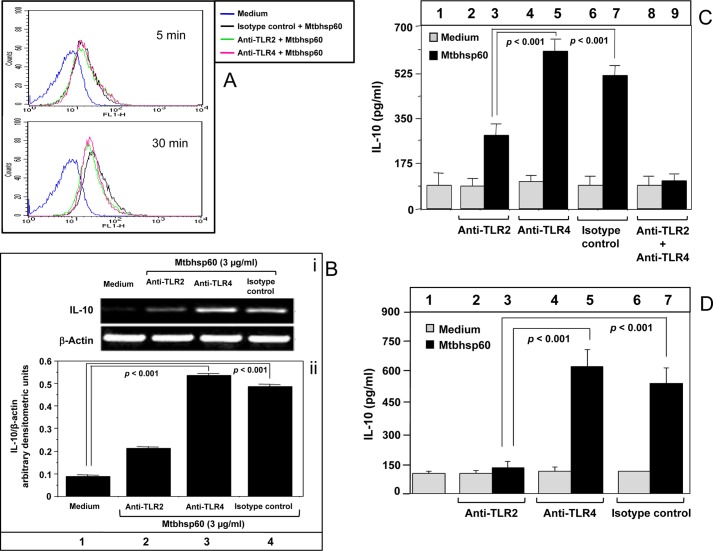
FIGURE 2.
IL-10 activation by Mtbhsp60 is TLR2-dependent. A, PMA-differentiated THP-1 macrophages were pretreated with either 10 μg/ml of anti-TLR2 mAb or anti-TLR4 mAb or isotype-matched control antibody for 1 h and then incubated with 10 μg/ml of biotin-labeled Mtbhsp60 at 4 °C for 5 and 30 min followed by incubation with streptavidin-FITC. The fluorescence was measured by flow cytometry. B, PMA-differentiated THP-1 macrophages were pretreated with 10 μg/ml of anti-TLR2 mAb or anti-TLR4 mAb or isotype-matched control antibody for 1 h and cultured for 2 h in the presence of 3 μg/ml of Mtbhsp60. Total RNA was extracted, and IL-10 levels were measured by semi-quantitative RT-PCR, and quantification of the IL-10 mRNA was performed by densitometric analysis using AlphaEaseFC software and the Spot Denso tool (version 7.0.1; Alpha Innotech, San Leandro, CA). Data are expressed as mean ± S.D. of three independent experiments. C, PMA-differentiated THP-1 macrophages were pretreated with neutralizing mAb to either TLR2 or TLR4, isotype-matched control antibody, or with both anti-TLR2 mAb and anti-TLR4 mAb in the absence or presence of Mtbhsp60 (3 μg/ml). After 48 h, IL-10 cytokine levels in culture supernatants from various groups were measured by EIA. D, experiments were also set to measure IL-10 levels by EIA in C57Bl/6 peritoneal macrophages treated with 10 μg/ml of anti-TLR2 Ab, anti-TLR4 Ab, or isotype control antibody in the absence or presence of Mtbhsp60 (3 μg/ml). Results shown are representative of three different experiments.

