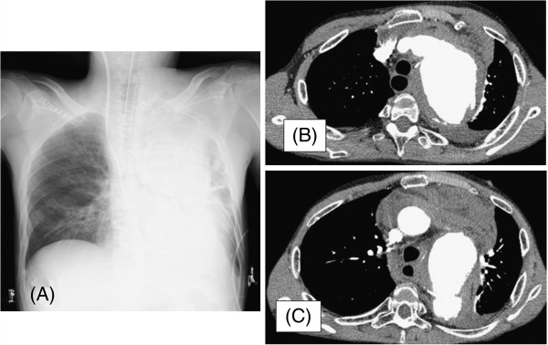
Figure 1.

Chest radiograph and computed tomography scan of patient with GPA. (A) Chest radiograph of a patient with a very large aneurysm of the aortic arch. Evident are marked widening of the mediastinum and aortic contour. (B) and (C) Axial contrast-enhanced computed tomography scan of the thorax revealing ruptured aortic aneurysm and collapsed left lung.
