
Figure 5.
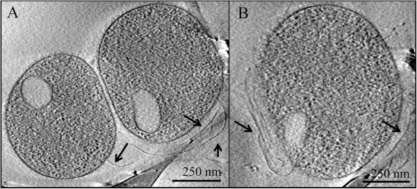
Cryo-electron microscopy of AMD plasma cells with putative pili. Panel A and panel B show evidence of pili on two different cells collected from the Richmond Mine AMD. Arrows point to pili. Vesicle-like structures are delineated by a single membrane layer around an ovoid shape in each cell’s cytoplasm.