Figure 5.
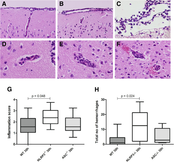
Histopathology in Asc−/−, Nlrp3−/−, and WT mice with pneumococcal meningitis. Representative brain slides showing neutrophil infiltration in the meninges 30 hours after induction of pneumococcal meningitis in a WT mouse (A), Asc−/− mouse (B) and Nlrp3−/− mouse (C). Perivascular cuffing 30 hours after induction of pneumococcal meningitis in a WT mouse (D), Asc−/− mouse (E) and Nlrp3−/− mouse (F), showing frequent intracerebral and subpial hemorrhages associated with neutrophil infiltration; meningeal, perivascular and intracerebral neutrophil influx was scored on a scale 0–4, means of 16 brain slides per mouse in the coronal plane are displayed (WT, n = 11; Asc−/−, n = 8; Nlrp3−/−, n = 9; G). Sum of intracerebral hemorrhages and subpial hemorrhages per mouse (WT, n = 11; Asc−/−; n = 8; Nlrp3−/−; n = 9; H).

