
Figure 1.
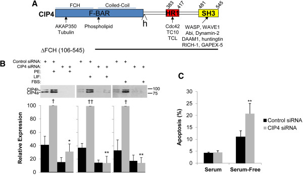
CIP4 structure and expression. A. Primary structure of CIP4 showing domains and binding sites for known protein partners. “h” indicates the 56 amino acid residue insertion present in CIP4h. B. Neonatal rat ventricular myocytes were transfected with control or CIP4 siRNA and then stimulated with 10 μM PE, 1000 U/mL LIF, or 10% FBS for two days as indicated. CIP4 proteins in whole cell lysates were detected using a mouse anti-CIP4 antibody. n = 4–5. †p-values vs. no drug control; *p-values vs. control siRNA-transfected myocytes treated with the same agonist. C. Myocytes were transfected with control or CIP4 siRNA and cultured in minimal media +/− 4% horse serum for two days before TUNEL staining. % TUNEL-positive nuclei are indicated. **p < 0.005 for CIP4 siRNA vs. control siRNA. n = 3–5.