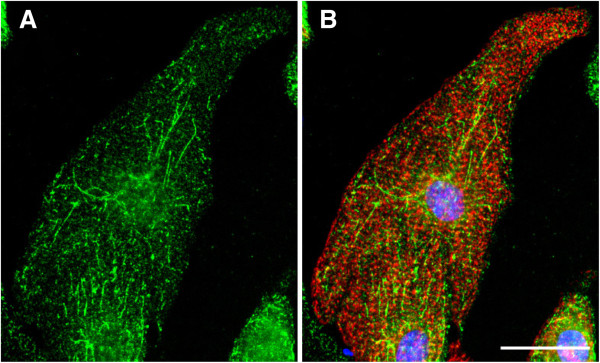
Figure 2.

CIP4 localization in myocytes. Myocytes expressing myc-CIP4 WT were stained with myc antibodies (green) and α-actinin (red) antibodies and Hoechst nuclear stain (blue). Bar = 20 μm. Panel A shows the green channel alone. n > 3. Panel B is a composite image of myc and actinin antibody staining and the nuclear stain.
