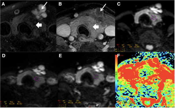
Figure 2.

A-36-year-female patient with thyroid papillary carcinoma at left lobe and isthmuses is shown. (A-B) Non-contrast and contrast transversal images showed abnormal signal at left lobe and isthmus with multiple cysts (long arrows). (C-E) showed ADC value measured from ADC map with b factors of 300, 500 and 800 s/mm2, respectively. (F) ADC map generated at b-factor of 300 s/mm2.
