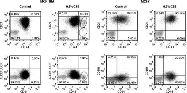
Figure 4.

CSE causes changes in stem cell markers in mammary epithelial cells and breast cancer cells. FACS analysis of MCF 10A cells treated with CSE for 30 weeks and MCF7 cells treated with CSE for 17 weeks. Cells labeled with isotype control antibodies, or incubated with DEAB (negative control) are shown in grey. Cells labeled with specific antibodies, or by aldefluor reaction are in shown in black and the percentage of cells in each quadrant is shown. MCF 10A: quadrants were established to include 99.9% of CD24+/CD44+, or ALDEFLUOR+/CD44+ mock treated cells; cells with increased CD44 positivity (circled) are concentrating in the lower right quadrants after CSE treatment, indicating loss of CD24 positivity and ALDEFLUOR signal. MCF7: quadrants were established to include 99% of signal from isotype antibody (negative); mock treated MCF7 cells consist of a mixed population of CD44+ and CD44- cells, uniformly CD24+; CSE treatment caused a shift to the CD24+/CD44+ quadrant, and an increase in CD49f+ cells.
