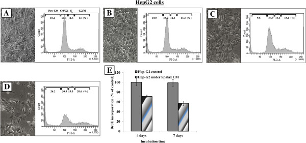
Figure 9.
Effects of Spalax, mouse and rat conditioned media on morphology and cell cycle progression in HepG2 cells. HepG2 cells were incubated under conditioned media for eight days; thereafter, cell morphology was documented using phase contrast microscopy, harvested, stained with PI and analyzed by flow cytometry. Representative images (×200) and flow cytometry histograms are presented: (A) control media; (B) rat CM; (C) mouse CM; (D) Spalax CM; (E) BrdU incorporation assay: HepG2 were grown in 96-well plates (2000 cells/well) for four and seven days under Spalax-generated CM. BrdU Cell Proliferation ELISA (Exalpha) was used. Time-dependent decrease in cell proliferation under Spalax-generated CM is depicted. CM, Conditioned media; PI, Propidium iodide.

