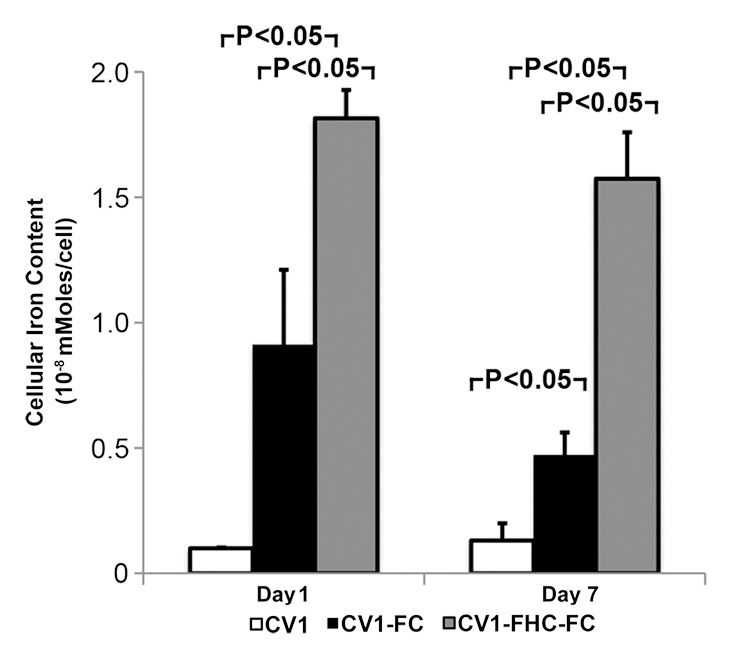
Figure 2d:

(a) Photomicrographs of dual immunofluorescent nuclear 4′,6-diamidino-2-phenylindole (DAPI) and human influenza hemagglutinin (HA) staining reveal robust perinuclear FHC expression in CV1-FHC fibroblasts. (b) Bar graph shows data for CV1-FC and CV1-FHC-FC cells imaged immediately after removal from FC-supplemented medium. CV1-FHC-FC cells showed significantly enhanced mean R2 contrast at increasing cell densities when compared with CV1-FC cells at day 1. Control (CV1) phantoms contained CV1 cells suspended at identical densities. * = P < .05 vs 1.0 ×10⁶ cells/mL, † = P < .05 vs 2.5 × 10⁶ cells/mL, ‡ = P < .05 vs 5.0 × 10⁶ cells/mL. (c) Bar graph shows that on day 7 after removal of FC-supplemented medium, mean R2 contrast remained significantly enhanced in CV1-FHC-FC compared with that in CV1-FC cells at increasing cell densities. * = P < .05 vs 2.5 ×10⁶ cells/mL, † = P < .05 vs 5.0 × 10⁶ cells/mL, ‡ = P < .05 vs 10 × 106 cells/mL. (d) Bar graph shows enhanced iron uptake on day 1 and retention on day 7 in CV1-FHC-FC cells.
