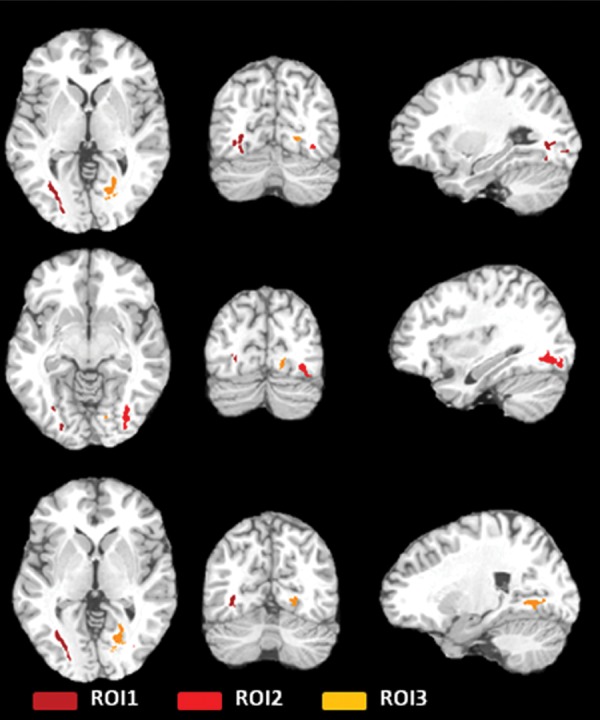
Figure 1b:

(a, b) Three ROIs in the temporo-occipital white matter detected by the initial voxelwise linear regression of estimated prior 12 months of heading on FA, shown as color regions rendered in three-dimensional images (a) and superimposed on T1-weighted axial (left), coronal (middle), and sagittal (right) images from the Montreal Neurological Institute template (b). FA at each ROI was significantly lower as a function of greater heading exposure. R = right.
