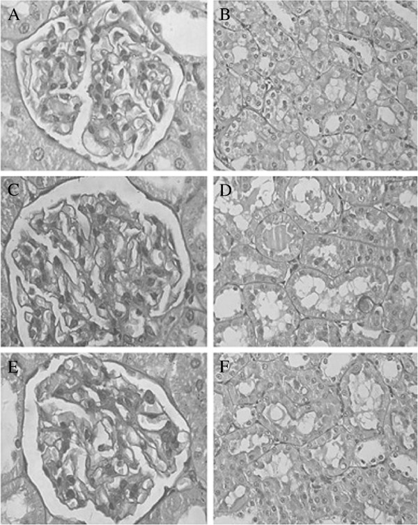
Figure 1.
Pathological pictures of rat kidneys from groups A, B, and C. (A) The normal renal glomerulus of group A (×400). (B) No obvious pathological change in renal tubules of group A (×400). (C, E) Mild glomerular mesangial proliferation in renal glomerulus of groups B and C (×400). (D, F) There were no obvious pathological changes in renal tubules in groups B and C (×400).

