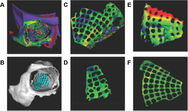
Figure 3.

3D shape analysis. (A) Manually superimposed pre- and post-operative virtual models – before matching procedure. (B) after matching procedure. (C) typical segmentation of a titanium mesh implant. (D) medial orbital wall as area of interest (green signifies no differences compared to the virtual planning). (E) orbital floor with transition zone to the medial wall (in red; differences are up to 1.5 mm). (F) complete titanium mesh implant shows an excellent result.
