
Figure 2.
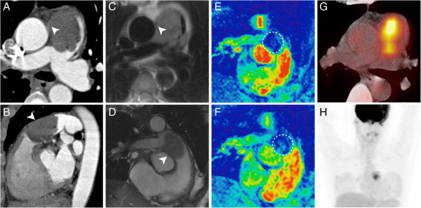
This figure illustrates an in-depth preoperative work-up in a 46-year-old female patient with an undifferentiated intimal sarcoma of the central pulmonary artery. Both, the MDCT (A, B) and the MRI (C, D) clearly demonstrate a subtotal occlusion of the pulmonary trunk due to an intraluminal process. Imaging findings are suggestive of a focal expansion beyond the vessel wall (A-D, arrowheads). The MR first-pass perfusion sequence (F) demonstrates a subtle perfusion of the lesion as compared to the non-enhanced control scan (E, circle). The F-18 FDG PET/CT proves a high metabolic activity (SUVmax = 7.8) within the lesion (G, H) and strengthens the suspicion of malignant disease.