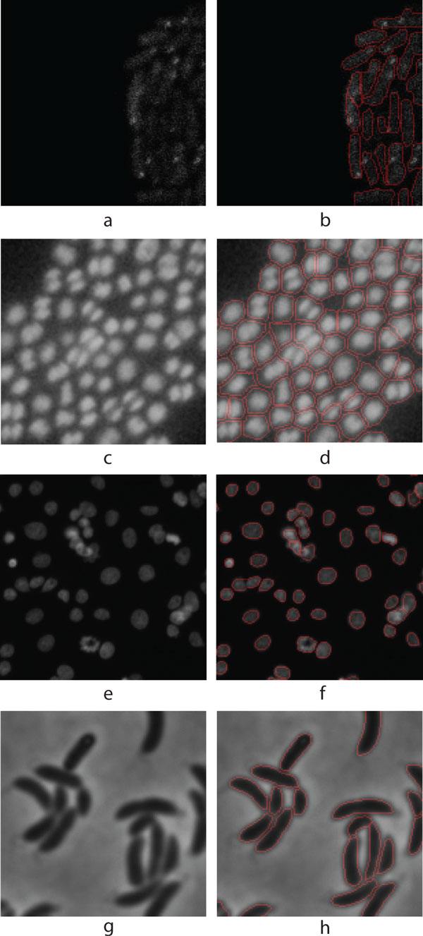
Figure 2.
Illustrative examples from the test samples. (a) Fluorescent protein labelled E. coli cells captured with confocal microscope, (b) Segmented result of (a), (c) Fluorescent protein labelled Staphylococcus cells in Epifluorescence microscopy image. (d) Segmented result of (c), (e) Human HT29 Colon Cancer 1 image set (Source [8]), (f) Segmented result of (e), (g) E. coli cells captured with phase contrast microscope (Source [11]), (h) Segmented result of (g).

