Figure 1.

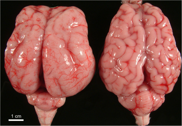
Affected and normal brains. Comparative image of a brain showing lissencephaly (left), with few and poorly developed sulci, and a marked cerebellar hypoplasia, and a brain from a newborn control lamb (right).

Affected and normal brains. Comparative image of a brain showing lissencephaly (left), with few and poorly developed sulci, and a marked cerebellar hypoplasia, and a brain from a newborn control lamb (right).