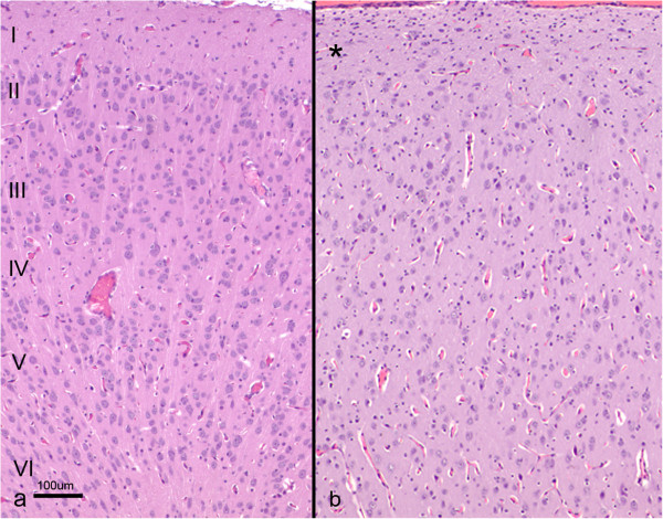
Figure 3.
Histological section of the cerebral cortex from a control (a) and lissencephalic (b) brain. Both sections were taken at the same magnification. Whereas in the control brain the whole cortex can be seen in the picture and organized in six layers (I to VI), in the lissencephalic brain only the most superficial part of the cortex is shown (aprox. 40% of the whole thickness), due to the thickening of this layer. A sparse-cellular layer underneath the piamater can be identified (*), whereas in the rest of the gray matter the neurons appear disorganized. H-E.

