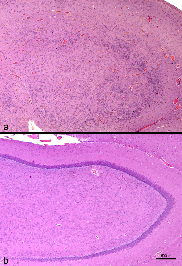
Figure 5.
Cross section of the hippocampus from a lissencephalic (a) and control (b) brain. Both sections were taken at the same magnification. Whereas in the control brain there is an evident and organized layer of pyramidal neurons, this region shows a marked cellular disorganization in the lissencephalic brain, with several layers of neuronal and glial cells interspersed. H-E.

