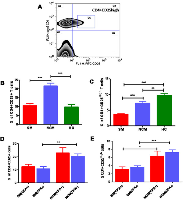
Figure 3.
Profile of CD4+CD25+ve cells in malaria patients with and without CFA. PBMCs of study subjects at the time of recruitment were analyzed by flow-cytometry for CD4 and CD25 expression after staining with anti-human CD4-PE.cy5 and anti-human CD25-FITC. Percentage of CD4+ve T cells expressing CD25 are shown here, A: Zebra plot for CD4+CD25high T–cells, G1 represents CD4+cells, G4 represents CD25+cells, and G3 represents CD4 + CD25+ cells and G5 represents CD4+CD25 high cells. Percentage of CD4+CD25+ T cells (B) and CD4+CD25high T (C) cells in SM=91, NCM=58 and control=22 are shown. The association within the groups was analysed by one way analysis of variance (ANOVA) followed by Tukey’s post-hoc test. Percentages of CD4+CD25+ve T cells and CD4+CD25high T cells in severe malaria (CFA+ve=14, CFA-ve=64), non-complicated malaria (CFA+ve=13, CFA-ve=34) patients with or without active filarial infection are shown in D and E respectively. The association within the groups were analysed by one way analysis of variance (ANOVA) followed by Tukey’s post- hoc test.

