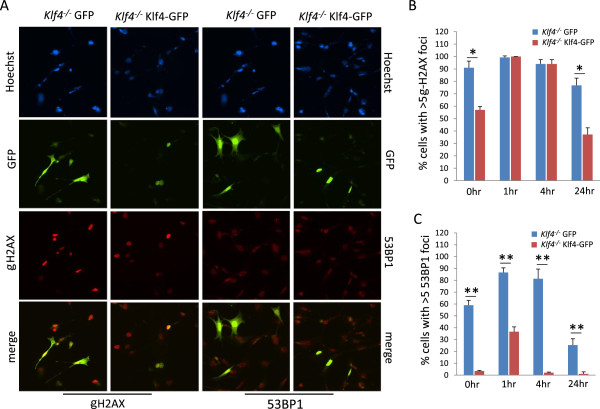
Figure 4.
Immunostaining for γ-H2AX and 53BP1 in Klf4−/−MEFs transfected with GFP or Klf4-GFP. (A) Immunostaining was conducted for γ-H2AX and 53BP1 in Klf4−/−MEFs transfected with GFP or Klf4-GFP.hoechst stain (blue) was used to visualize nuclei. Shown is a representative result of three independent experiments for the non-irradiated cells. (B) Histogram showing quantification of cells with ≥ 5 γ-H2AX foci in Klf4−/−MEFs cells transfected with GFP or Klf4-GFP, with or without γ-irradiation. (C) Histogram showing quantification of cells with ≥ 5 53BP1 foci in Klf4−/−MEFs cells transfected with GFP or Klf4-GFP, with or without γ-irradiation. For (B) and (C), foci were counted for non-irradiated cells, and at 1, 4 and 24 h post irradiation. One hundred green cells were counted per cell type per experiment. N = 5; * p < 0.05, ** p < 0.01 compared to GFP-transfected Klf4−/− cells.

