Figure 3.
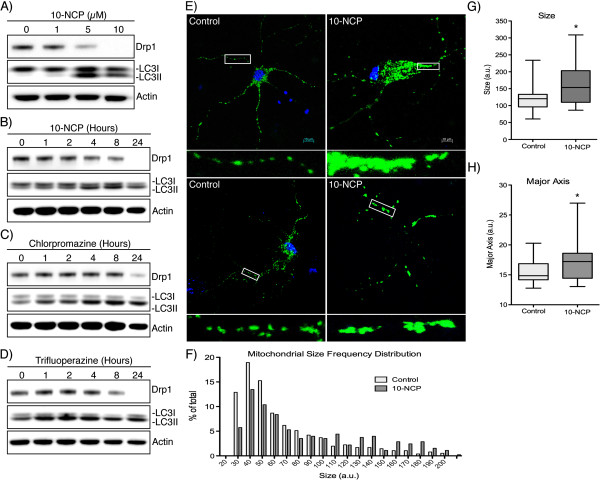
Inducing autophagy decreases Drp1 levels in rat-derived striatal neurons. (A) E18 rat-derived striatal neurons were incubated with 10-NCP at the concentrations indicated for 12 hours. (B) Neurons were incubated with 5 μM 10-NCP for the indicated times. (C and D) Chlorpromazine and Trifluoperazine (5 μM) was incubated for the times indicated followed by Western blotting. (E) Striatal neurons were incubated with 10-NCP for 12 hours along with transduction of CellLight Mitochondria GFP to allow labeling and visualization of mitochondria. (F) Frequency distribution of mitochondrial size in control and 10-NCP treated neurons as quantitated from confocal immunofluorescent images using the Mitochondrial Morphology plug-in for ImageJ (n > 500 for each condition, X-axis truncated at 200). (G and H) Box and whisker plot (with whiskers representing 5-95th percentile) of the average mitochondrial size and major axis per neuron (n = 20). *P < 0.05.

