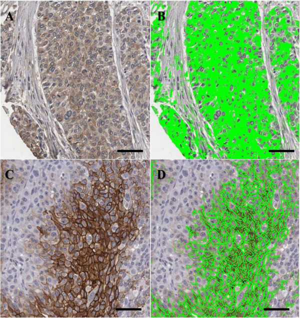
Figure 2.
Digital image analysis of cytoplasmic and membranous staining. Cytoplasmic HIF-1α staining is shown (A) and automated image analysis utilizing TissueIA recognizes cytoplasmic HIF-1α staining highlighted in green color (B). CA9 is shown in membranous staining (C) and automated image analysis determines membranous CA9 staining highlighted in green color (D). The output from the algorithm returns a number of quantitative measurements for intensity and percentage of positive staining present. Scale bar: 100 μm.

