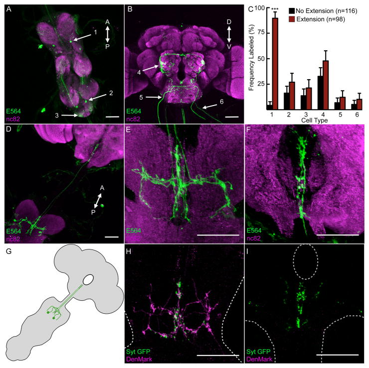
Figure 2. Mosaic analysis identifies a single cell-type responsible for constitutive proboscis extension.
A-B. Expression of UAS-mCD8-GFP in E564-Gal4 neurons in VNC (A) and central brain (B). The six neural types in mosaics are labeled (arrows). See Figure S2 for single-cell clones of the six different neural types.
C. Flies expressing Kir2.1 and CD8-GFP in subsets of E564 neurons were assayed for constitutive proboscis extension (extension, red bars, n=98), or normal proboscis posture (no extension, black bars, n=116) (genotype: tub>Gal80>; E564-Gal4,UAS-mCD8::GFP/UAS-Kir2.1; MKRS, hs-FLP flies). The frequency of the 6 cell-types identified in A and B is shown in extenders and non-extenders. All cell-types except for cell-type #1 showed similar distribution in both groups. mean±95% CI, Fisher’s exact test, *** P<0.001.
D. A clone of cell-type #1 (PERin) expressing mCD8-GFP, showing cell bodies in the first thoracic segment and projections to the SOG.
E-F. Detailed image of PERin clone from D in first thoracic segment (E) and SOG (F).
G. Schematic of PERin showing processes in the VNC and SOG.
H-I. E564-Gal4 line expressing the presynaptic marker UAS-Syt-GFP and the postsynaptic marker UAS-DenMark. Images are the same regions shown in E, F (PERin), indicating mixed pre-and postsynaptic fibers in the first thoracic segment (H), and presynaptic fibers in the SOG (I). The edges of the VNC (H) and SOG (I) are shown with dotted lines. Scale bars are 50μm. See Figure S2 and Videos S1 and S2 for anatomical studies showing that PERin does not come into proximity of gustatory sensory dendrites or proboscis motor axons.

