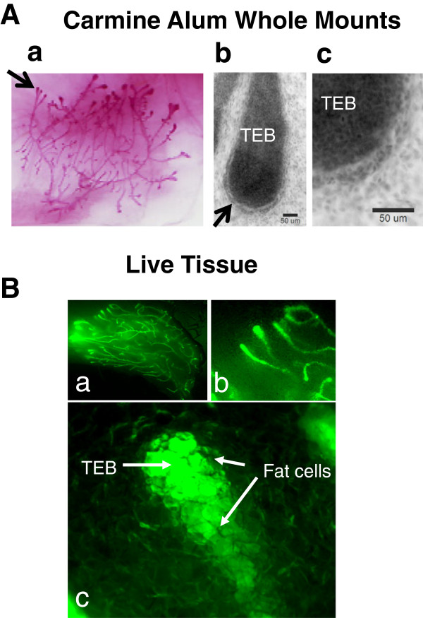
Figure 1.
Stereofluorescence microscopy of live mouse mammary gland, ex vivo. GFP-mice were sacrificed and the 3rd inguinal gland was excised. A a-c. Carmine Alum-stained glands used for multiphoton imaging of normal TEBs in this study (see Figures 4, 8, and 11) were imaged using bright field optics, first with a Nikon SMZ1500 stereofluorescence (A a) and then using a Nikon E600 upright microscope (A b, Nikon 20X/ 0.5 N.A., and c, Nikon 40 X /0.95 N.A.). Scale bars = 50 μm. B a-c. Another mammary gland from a GFP-mouse was imaged using the stereofluorescence microscope. Fat cells, just visible in B a-b at lower magnifications, are viewed surrounding the ductal epithelium obscuring cellular details of the terminal end bud (TEB) in B c.

