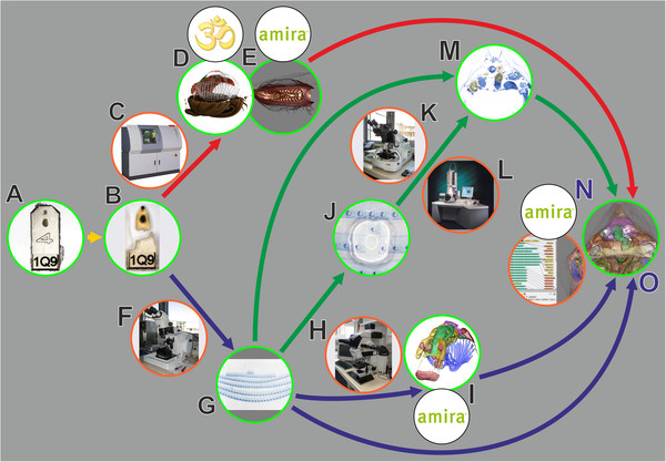
Figure 1.
General workflow for combining microCT, LM and TEM. Red arrows, volume data from microCT; blue arrows, LM section data; green arrows, TEM section data; green encircled, material and (interim) results; orange encircled, microscopic equipment; black encircled, software systems. A. EM fixed specimen embedded in epoxy resin block, anterior one used in study. B. Block trimmed and anterior portion detached, after microCT scan. C. MicroCT. D. Volume rendering with Drishti. E. Volume rendering with AMIRA®. F. Microtome for LM sectioning. G. LM section series. H. Light microscope. I. Surface rendering with AMIRA®. J. Remounting of LM sections for ultrathin sectioning. K. Ultramicrotome. L. TEM. M. 2D alignment for figure plates. N. 3D registration with AMIRA®. O. Combined 3D visualization of all datasets.

