Figure 2.

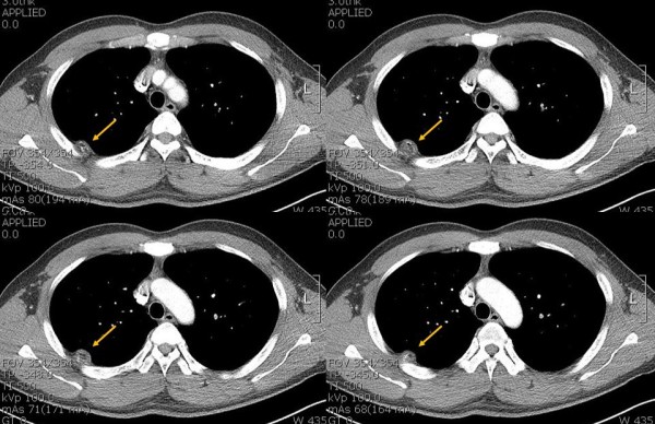
Contrast-enhanced computed tomography (CT) demonstrates the intrathoracic fatty mass with calcification that has obtuse margin projecting into the right hemithorax (arrow).

Contrast-enhanced computed tomography (CT) demonstrates the intrathoracic fatty mass with calcification that has obtuse margin projecting into the right hemithorax (arrow).