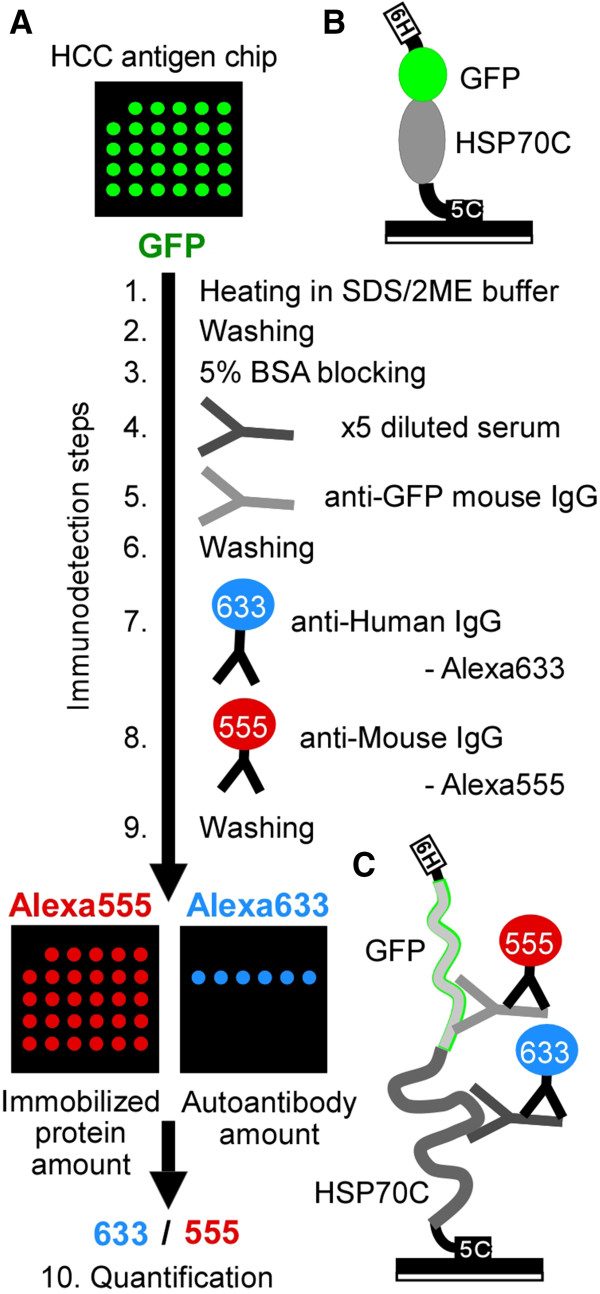
Figure 1.

Autoantibody detection on chip. (A) Autoantibody detection procedures. Protein chips were immersed in SDS/2ME sample buffer and heated at 95°C for 5 min (Immunodetection step 1) and washed (step 2). The chips were blocked with BSA (step 3), incubated with diluted serum (step 4) or mouse anti-GFP monoclonal IgG (step 5), washed (step 6), incubated with AlexaFluora 633 (Alexa633)-conjugated anti-human IgG to quantify bound serum autoantibodies (step 7), incubated with AlexaFluore 555 (Alexa555)-conjugated anti-mouse IgG antibody to quantify the amount of GFP-tagged protein (step 8), and washed (step 9). The chips were examined for Alexa555 and Alexa633 fluorescence of the same spot in the same chip and Alexa633 fluorescence of each spot was normalized using Alexa555 fluorescence of the same spot (step 10).(B-C) Schema of predicted protein chip surface before step 1 (B) and after step 9 (C). 6H: 6xHis-tag, 5C: 5xCys-tag.
