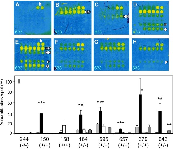
Figure 7.
Autoantibodies detection by the third-designed protein chip. (A-H) Full color autoantibody-detected images by ×10 diluted sera and anti-human IgG-Alexa633. The representative chip images from four serum samples tested in Figure 5, No. 244 (A), No.150 (B), No. 158 (C), No. 164 (D) and three new HCV+/HCC + serum samples, No. 595 (E), No. 657 (F), 679 (G), and a HCV+/HCC- serum sample, No. 643 (H), are shown. (I) Quantitative results of the third-designed chip. Others are shown same as Figure 6.

