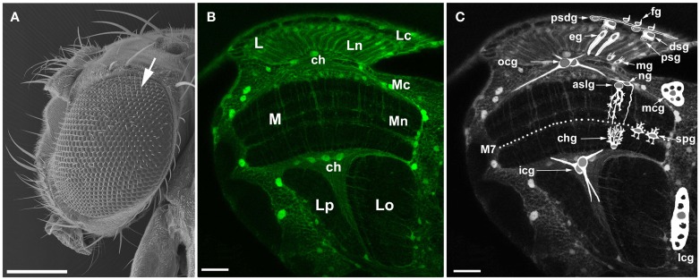
Figure 1.
The visual system of the fruit fly, Drosophila melanogaster. (A) Scanning electron micrograph of a head and a large compound eye. The eye is composed of approximately 800 hexagonal units called facets or ommatidia (arrow). The ommatidial array of photoreceptors in the retina receives photic and visual information, transduces it into receptor action potentials and transmits to underlying optic lobe. Scale bar: 200 μm. (B) Confocal image of the optic lobe of transgenic flies Repo-Gal4 × UAS-S65T-GFP, in horizontal section. Targeted expression of Green Fluorescence Protein (GFP) to glial cells reveals the general morphology of the optic lobe. There are three synaptic regions (neuropils) beneath the retina of the compound eye: the lamina (L), the medulla (M), and the lobula that in Diptera consists of the lobula (Lo) and the lobula plate (Lp). Lc, lamina cortex; Ln, lamina neuropil; Mc, medulla cortex; Mn, medulla neuropil; ch, chiasm. Scale bar: 20 μm. (C) Schematic representation of so far identified types of glia (based on Edwards et al., 2012) revealing their general morphology and relative locations in the optic lobe: fg, fenestrated glia; psdg, pseudocartridge glia; dsg, distal satellite glia; psg, proximal satellite glia; eg, epithelial glia; mg, marginal glia; mcg, medulla cortex glia; aslg, astrocyte-like glia of the distal medulla neuropil; ng, another type of the distal medulla neuropil glia; spg, serpentine glia; chg, chandelier glia; ocg, outer chiasm glia (giant and small ocg); icg, inner chiasm glia; lcg, lobula cortex glia; M7, the serpentine layer. Certain types of glia (eg, aslg, and/or ng, mcg, lcg, ocg, icg) can be also discern in the tissue visible in the background being marked by GFP. Scale bar: 20 μm.

